|
|
||||||||||||||||||||||||||||||||||||||||||||
|
Return to Glyco Navigation Page
|
||||||||||||||||||||||||||||||||||||||||||||
|
Glycopeptide Heterogeneity and Mass Pattern Recognition
|
||||||||||||||||||||||||||||||||||||||||||||
|
|
||||||||||||||||||||||||||||||||||||||||||||
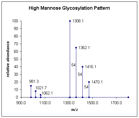
Example ESI Mass Spectrum
Above is a sample electrospray mass spectrum showing peaks that differ by 54 amu this is typical of a glycopeptide displaying mannose type glycosylation heterogeneity at the [M+3H]3+ charge state (see the chart above). To confirm the observation look for peaks that differ by 40.4 amu in the lower end of the spectrum indicating a quadruply charged series.
|
||||||||||||||||||||||||||||||||||||||||||||
| Simple monosaccharide mass table | Low mass CID marker ions | |||||||||||||||||||||||||||||||||||||||||||
|
References IUPAC-IUB Joint Commission on Biochemical Nomenclature (JCBN), Nomenclature of glycoproteins, glycopeptides and peptidoglycans glycopeptides and peptidoglycans glycopeptides and peptidoglycans, Recommendations 1985. |
||||||||||||||||||||||||||||||||||||||||||||
|
Return to Glyco Navigation Page home
| disclaimer |
||||||||||||||||||||||||||||||||||||||||||||
